Researchers at the Hebrew University of Jerusalem, Tel Aviv Sourasky Medical Center and Shaare Zedek Medical Center, have developed a new MRI technology that non-invasively assesses iron levels in the human brain, shedding light on its critical role in brain function, aging, and diseases like cancer and neurodegenerative disorders. This innovative approach, based on quantitative MRI, distinguishes between healthy and pathological brain tissue without contrast agents and provides insights into iron-related disorders, offering a significant advancement in neuroscience and healthcare.
Jerusalem, Israel – [September 12, 2023] – Research team led Shir Filo and Prof. Aviv Mezer of Hebrew University of Jerusalem and Dr. Tal Shahar currently, Director of the Neurosurgical Oncology Unit at the Neurosurgery Department, Tel Aviv Sourasky Medical Center, have unveiled a groundbreaking magnetic resonance imaging (MRI) technology that promises to revolutionize our understanding of iron homeostasis in the human brain. Their research, demonstrates the ability to non-invasively assess different molecular iron environments within the brain, shedding light on its vital role in normal brain function, aging, neurodegenerative diseases, and cancer.
Traditional MRI scans provide qualitative images that necessitate subjective interpretation by medical professionals. In contrast, quantitative MRI seeks to replicate the precision of assessing body temperature accurately when ill, moving beyond qualitative observations like “too hot” or “too cold.” Achieving this involves intricate physical models that amalgamate multiple MRI scans to extract a diverse array of measurements. These MRI measurements are then leveraged to uncover valuable biological insights, akin to a blood test’s ability to reveal our blood’s composition, encompassing proteins, fats, and potential irregularities. One critical aspect of brain function is maintaining the balance, or homeostasis, of iron, crucial for overall health. Imbalances in brain iron levels have implications for various conditions, including neurodegenerative diseases and cancer. Until recently, the non-invasive assessment of the molecular iron environment within the living human brain posed a significant challenge.
The research team from Hebrew University of Jerusalem, Tel Aviv Sourasky Medical Center and Shaare Zedek Medical Center introduces a novel MRI approach based on the quantitative MRI, which can sensitively detect changes in brain iron homeostasis. In their in vitro experiments, this MRI technology revealed distinct paramagnetic properties of key iron compounds, including ferritin, transferrin, and ferrous iron. The real breakthrough came with in vivo MRI scans of patients with brain tumors, operated at the Neurosurgery Department at Shaare Zedek Medical Center where the technology was also validated against ex vivo iron compound quantification of the resected tumors. These ex-vivo transcriptomics and proteomics analyses were done in the lab of Dr. Naomi Habib in the Hebrew University and the lab of Molecular Neuro-oncology at Shaare Zedek Medical Center, directed at the time of the study by Dr. Tal Shahar.
This innovative MRI approach demonstrates sensitivity to changes in iron mobilization capacity across different brain regions and during brain aging. It offers insights into changes in iron homeostasis and iron-related gene expression in pathological tissues. Most notably, it can differentiate between tumor tissue and non-pathological tissue without the need for toxic contrast agents injection.
Shir Filo, explained, “Our technology opens up new possibilities for understanding the role of iron in cancer, normal aging, and neurodegenerative diseases. It enables non-invasive research and diagnosis of iron homeostasis in the living human brain, offering a potential game-changer for healthcare and neuroscience.”
Prof. Aviv Mezer emphasized, “The strength of our team lies in its diverse composition, comprising experts from various disciplines and specialties. Dr. Tal Shahar was leading the medical aspect of the study involving meningioma patients and the proteomic studies. Dr. Rona Shahrabani was instrumental in conducting experiments with iron proteins, while Dr. Nevo Margalit, Chair of the Neurosurgery Department at Shaarei Zedek Medical Center made a substantial contributions through his involvement in a significant portion of the surgeries and recruitment of patients. Additionally, Dr. Eli Ben David, a radiologist at Shaarei Zedek Medical Center, played an indispensable role in enabling the MRI scans and Dr. Naomi Habib brought the valuable knowledge in gene expression. Their collective and truly interdisciplinary expertise and unwavering dedication significantly enhanced the success of this pioneering study.”
 In summary, the Hebrew University of Jerusalem’s MRI quantification, represents a groundbreaking advance in our ability to assess iron homeostasis in the human brain. It shows promising ability to distinguish between pathological and healthy brain tissue without the use of toxic contrast agents and offers insights into molecular iron states inaccessible to conventional MRI techniques. The technology’s sensitivity to in vitro, in vivo, and ex vivo iron environments validates its potential to enhance our understanding of iron-related disorders and opens new avenues for research and diagnosis in the living human brain.
The study, titled “ Non-invasive assessment of normal and impaired iron homeostasis in the brain,” has been published in Nature Communications. DOI for the paper is 10.1038/s41467-023-40999-z
Date of publication: September 12th 10:00 London Time – 05:00 ET
Research Team: Shir Filo-1, Rona Shaharabani-1, Daniel Bar Hanin-1, Miriam Adam-1, Eliel Ben-David-2, Hanan Schoffman-3, Nevo Margalit-4, Naomi Habib-1, Tal Shahar3,4,5, Aviv A Mezer-1
Institutions:
1-The Edmond and Lily Safra Center for Brain Sciences, The Hebrew University of Jerusalem, Israel
2-The Department of Radiology, Shaare Zedek Medical Center, Faculty of Medicine, The Hebrew University of Jerusalem, Israel
3-The Laboratory of Molecular Neuro-Oncology, Shaare Zedek Medical Center, Faculty of Medicine, The Hebrew University of Jerusalem, Israel
4-The Department of Neurosurgery, Shaare Zedek Medical Center, Faculty of Medicine, The Hebrew University of Jerusalem, Israel
5-The Department of Neurosurgery, Tel Aviv Sourasky Medical Center, Affiliated with Sackler Faculty of Medicine, Tel Aviv University, Tel Aviv, Israel
Pictures – all the pictures can be credited to the authors of the paper.
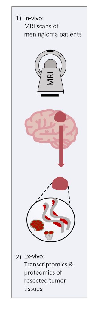
picture 1: A summary of the new non-invasive approach for assessment of normal and impaired iron homeostasis in the brain.
picture 2: The new MRI technique was validated by comparing in vivo MRI scans of meningioma patients with ex vivo iron quantification of the patiens’ tumor tissues.
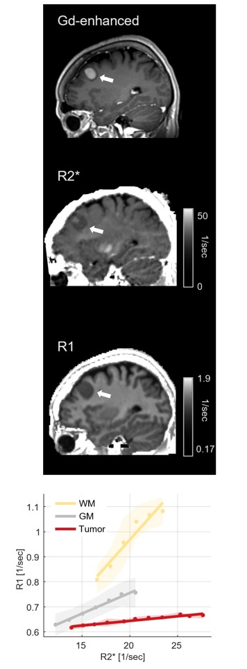
picture 3: : MRI image with contrast agent (Gd-enhanced), and quantitative MRI images (R1 and R2*) in a representative subject with a meningioma brain tumor (white arrow). Our new approach (in the bottom) separates between healthy tissue (WM and GM) and tumor tissue.
The Hebrew University of Jerusalem is Israel’s premier academic and research institution. With over 25,000 students from 90 countries, it is a hub for advancing scientific knowledge and holds a significant role in Israel’s civilian scientific research output, accounting for nearly 40% of it and has registered over 11,000 patents. The university’s faculty and alumni have earned eight Nobel Prizes and a Fields Medal, underscoring their contributions to ground-breaking discoveries. In the global arena, the Hebrew University ranks 86th according to the Shanghai Ranking. To learn more about the university’s academic programs, research initiatives, and achievements, visit the official website at http://new.huji.ac.il/en